Services

Credibly reintermediate backend ideas for cross-platform models. Continually reintermediate integrated processes through technically sound intellectual capital.
COVID-ICU-illustration.jpg

Intensive care units (ICUs)

What is an intensive care unit (ICU)?

Intensive care refers to the specialised treatment given to patients who are acutely unwell and require critical medical care.

An intensive care unit (ICU) provides the critical care and life support for acutely ill and injured patients.

Unless you are an emergency admission, you will need a referral from your doctor or specialist to be admitted to ICU.

Who is cared for in ICU?

Patients may have a planned admission following surgery, an unexpected admission after an accident or be admitted because of a sudden and critical deterioration to their health.

ICU teams are multi-disciplinary, made up of highly skilled intensive care nurses, doctors and specialists trained in providing critical care for patients with a variety of medical, surgical and trauma conditions.

Some hospital ICUs specialise in providing care for particular health conditions or injuries including:

  • major trauma
  • severe burns
  • respiratory failure
  • organ transplants
  • complex spinal surgery
  • cardiothoracic surgery.

What to expect in ICU

ICU is one of the most critically functioning operational environments in a hospital.

Every ICU in a hospital has a different environment that will reflect the specialist medical and surgical procedures they perform.

Most ICUs are fairly large sterile areas with a high concentration of specialised, technical and monitoring equipment needed to care for critically ill patients.

The ICU environment can be confronting for some patients and visitors who may find the activity, sounds, machines, tubes and monitors intimidating.

When you visit someone you care about in ICU it can be an uncomfortable experience –  you may feel helpless, overwhelmed, frustrated and sad. Your feelings and apprehension are understood by the staff that provide support for the people you care about.

Typically ICU also has a higher ratio of doctors and nurses to patients.

ICU equipment

It can be a frightening and uncertain time for you, family and friends to see people you care about being monitored and supported by machines.

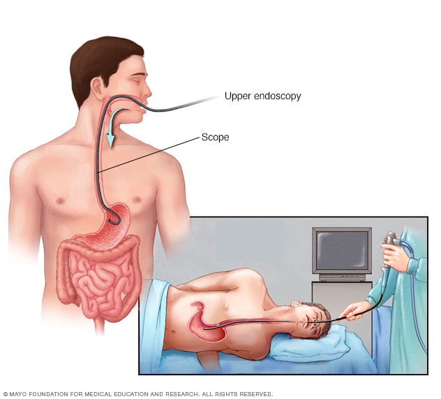
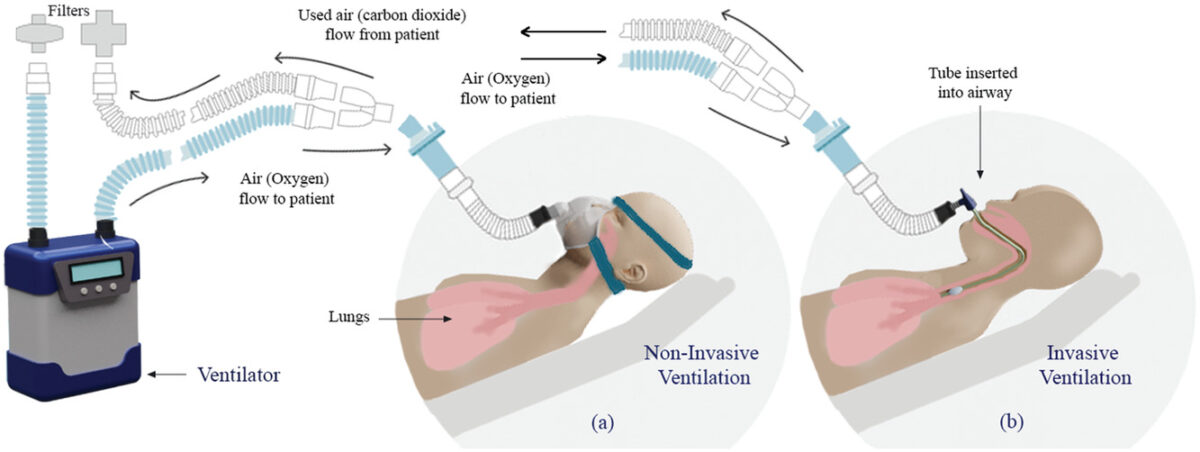
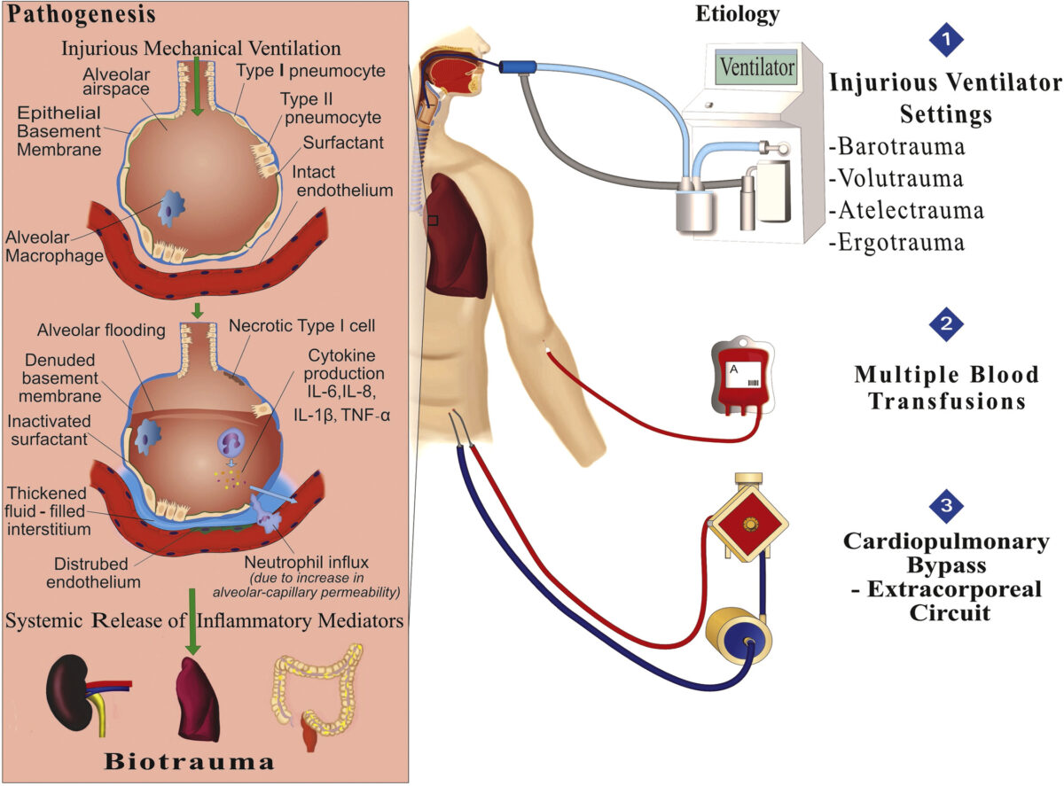
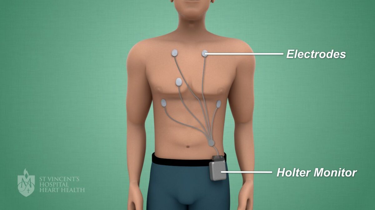
In ICU you will see many patients connected to a heart monitor, others will be supported with breathing assistance from artificial ventilators, be on dialysis machines and receiving a variety of intravenous infusions via tubes and drips.

Be prepared to see lots of lines, tubes, wires and monitoring equipment. Almost all ICU equipment uses alarms to let staff know about a change in a patient’s condition. Not all equipment alarms signal an emergency situation.

Visitors

Every ICU has a visitor policy to ensure the wellbeing of their patients. You will need to ask ICU hospital staff about their specific visiting hours and requirements.

Visiting is usually restricted to people the patient considers to be immediate family.

If you are feeling unwell or have an existing health condition you should reconsider visiting ICU or discuss your circumstances with ICU staff before you plan to visit.

Hygiene

As intensive care patients are very vulnerable to infections, it is important that visitors wash their hands before entering ICU to prevent transferring infection.

Mobile phones

Mobile phones should be turned off in ICU as they may interfere with vital electrical equipment supporting patients.

Gifts

Restrictions are in place to allow easy access to vital medical equipment and to patients.

If you are unsure what you can bring with you check with ICU staff before you plan to visit.

Patient and family support services

Counselling

Admission to ICU because of critical illness or accident can have a huge physical and emotional impact on your life and your family.

Some ICUs have a dedicated counsellor to provide support for patients and their families. These counsellors are highly experienced and have a thorough knowledge of ICU procedures.

Hospital counselling support services are also available at all major hospitals.

Pastoral care

For many people, emotional and spiritual thoughts tend to surface when someone they care about is in a critical condition in hospital.

Many hospitals provide chaplaincy and pastoral counselling services for patients, families and staff who need compassionate, professional and spiritual guidance and support.

Some hospitals also have a non-denominational chapel available for times of reflection and prayer.

Interpreter service

An interpreter service is available for patients and families if English is not your first language.

These interpreters are specifically trained to interpret medical terms into other languages. It is important that you use this service if you are having problems understanding doctors explaining information or are being asked to provide consent for medical procedures.

Speak to ICU staff if you would like to use interpreting services.

mcdc7_endoscopy-8col.jpg

Overview

An upper endoscopy is a procedure used to visually examine your upper digestive system with a tiny camera on the end of a long, flexible tube. A specialist in diseases of the digestive system (gastroenterologist) uses an endoscopy to diagnose and, sometimes, treat conditions that affect the esophagus, stomach and beginning of the small intestine (duodenum).

The medical term for an upper endoscopy is esophagogastroduodenoscopy. You may have an upper endoscopy done in your doctor’s office, an outpatient surgery center or a hospital.


Why it’s done

An upper endoscopy is used to diagnose and, sometimes, treat conditions that affect the upper part of your digestive system, including the esophagus, stomach and beginning of the small intestine (duodenum).

Your doctor may recommend an endoscopy procedure to:

  • Investigate symptoms. An endoscopy may help your doctor determine what’s causing digestive signs and symptoms, such as nausea, vomiting, abdominal pain, difficulty swallowing and gastrointestinal bleeding.
  • Diagnose. Your doctor may use an endoscopy to collect tissue samples (biopsy) to test for diseases and conditions, such as anemia, bleeding, inflammation, diarrhea or cancers of the digestive system.
  • Treat. Your doctor can pass special tools through the endoscope to treat problems in your digestive system, such as burning a bleeding vessel to stop bleeding, widening a narrow esophagus, clipping off a polyp or removing a foreign object.

An endoscopy is sometimes combined with other procedures, such as an ultrasound. An ultrasound probe may be attached to the endoscope to create specialized images of the wall of your esophagus or stomach. An endoscopic ultrasound may also help your doctor create images of hard-to-reach organs, such as your pancreas. Newer endoscopes use high-definition video to provide clearer images.

Many endoscopes allow your doctor to use technology called narrow band imaging, which uses special light to help better detect precancerous conditions, such as Barrett’s esophagus.

gch2202000046-fig-0003-m-1200x452.jpg

NIV refers to the provision of respiratory support without direct tracheal intubation. As such, it aims to avoid some of the complications inherent with invasive ventilation, such as the need for sedation with risks of hemodynamic instability and subsequent risk of delirium, nosocomial infection, etc.1 In a recent worldwide survey, the use of NIV increased from 4% to 11% of all episodes of mechanical ventilation between 2001 and 2004 respectively, with even higher rates of use in some European countries.2 Indeed, for some situations it is considered the first-choice mode of ventilatory support However, it may not be appropriate for all patients, particularly as it is being used increasingly outside of the traditional critical care setting (Indications/Contraindications for NIV).


gr1_lrg-1200x887.jpg

Introduction

Indications for ventilation

Early identification of critically ill patients, before the occurrence of significant cardio-respiratory decompensation, is one of the major goals of critical care outreach programmes. Patients who require ventilatory support often develop a common pattern of physiological deterioration including:

  • increasing respiratory rate
  • asynchronous respiratory pattern
  • a change in mentation and level of consciousness
  • frequent oxygen desaturation despite increasing oxygen concentration
  • hypercapnia and respiratory acidosis
  • circulatory problems, including hypotension and atrial dysrhythmias.


Modern ventilators

The modern ventilator is a complex computer-driven tool and a detailed description of its modes and use are beyond the scope of this article (see Further reading). In simple terms, it mixes air under pressure with variable oxygen concentrations to provide inspiration and expiration, each ‘breath’ is characterized by three factors, which can be adjusted by the operator;

  • Trigger: the ventilator can deliver a breath according to a timer that defines a specific set rate (ventilator initiated/mandatory breaths), or as a result of the patient’s own breathing efforts effecting a change in the pressure or flow in the ventilator circuit (patient-initiated/spontaneous breaths).
  • Target: the flow of air into the lung can be to a specific target flow rate (volume control) or pressure (pressure control; pressure support; bi-level)
  • Termination: the signal for the ventilator to finish inspiration and allow expiration (passive) may be the achievement of a specific volume (volume-cycled: volume control), after a specific time (time-cycled: pressure control/bi-level) or following the reduction of inspiratory flow to a preset level (flow-cycled: pressure support)

index.jpg

Benefits of Treatment

Peripheral angioplasty and stenting open a narrowed or blocked artery in your extremities, like your arms or legs. This treatment can prevent the need for more invasive procedures, such as bypass surgery.

Your doctor may suggest angioplasty and stenting if you have peripheral artery disease (PAD), which can cause:

  • Chronic pain in your legs.
  • Heavy feeling in your legs.
  • Limitations to your daily activities.
  • Wounds on your legs or feet that do not heal.

Before Your Angioplasty and Stenting Procedure

Before your procedure, your vascular surgeon will conduct a thorough exam.

He or she will use imaging tests — like ultrasound or a CT scan — to find the blockages and see how severe.

What to Expect During Peripheral Angioplasty and Stenting

During your procedure, your surgeon will:

  1. Insert a tiny needle into the blood vessel in your groin or upper arm.
  2. Thread a soft guidewire through the needle and advance it to the right location while watching it with live x-ray.
  3. Inject a dye through the needle that aids in finding the arterial blockage(s).
  4. Insert a small device that looks like a tiny balloon inside your artery.
  5. Inflate the balloon and compress the plaque outward on the walls of the artery to opens it, allowing for better blood flow.
  6. Insert a stent — a mesh tube made of metal. The stent serves as a permanent metal scaffold to keep the artery open.

Depending on your condition and overall health, you may be able to go home the same day. Most people spend one day or less in the hospital.

10_HolterMonitor-1200x675.jpg

What is a holter monitor?

A holter monitor is a device that records your heart beats. It contains electrodes, wires and a small machine that attaches to your waistband.


Why do I need a holter monitor?

You may need a holter monitor if your doctor thinks there could be a problem with your heart rate or rhythm. You might have symptoms such as:

  • Fainting
  • Dizziness
  • Shortness of breath
  • Chest pain
  • Irregular heartbeat

Even if you’ve had an ECG you may still need to wear a holter monitor. ECGs only monitor your heartbeat for a short time – which isn’t always long enough to identify potential problem.

What are the risks of having a holter monitor?

Holter monitoring is a safe and painless test with no significant risks. Some people experience minor skin irritation from the sticky dots (electrodes) used to monitor the heartbeat.

How do I prepare for a holter monitor?

There’s not a lot you need to do to prepare – but you will need to shower. After your holter monitor has been fitted, you can’t get it wet or remove it. The hair on your chest may need to be shaved to help the electrodes stick to your skin.

What happens during a holter monitor?

The holter monitor test is a very straightforward procedure:

  • Electrodes are stuck to your chest
  • The electrodes are connected to the small holter machine device
  • You’ll be shown how to attach the device to your clothes

While the device is fitted, you’ll need to:

  • Wear it during your usual activities
  • Keep it on at all times, even during sleep
  • Never take it off
  • Keep a diary of all your daily activities and the time you did them
  • Record any specific symptoms you feel (dizziness, light-headedness, chest pain, shortness of breath)

What happens after a holter monitor?

After one or two days, you’ll return to your doctor’s office with your diary. Your doctor will remove the holter monitor, discuss your results and recommend an appropriate treatment plan to help you manage your symptoms moving forward

jcm-08-01176-g001.jpg

Electrical impedance tomography (EIT) is a radiation-free functional imaging modality that allows non-invasive bedside monitoring of both regional lung ventilation and arguably perfusion. Commercially available EIT devices were introduced for clinical application of this technique, and thoracic EIT has been used safely in both adult and pediatric patients

Electrical resistivity of thoracic tissues.

Tissue Resistivity (Ω·cm)
Blood 150
Lungs, inspiration 2400
Lungs, expiration 700
Heart muscle, longitudinal 125
Heart muscle, transversal 1800
Skeletal muscle, longitudinal 160–575
Skeletal muscle, transversal 420–5200
Fat 2000–2700
Bone 16,600

3. EIT Measurements and Image Reconstruction

Electrodes are either single self-adhesive electrodes (e.g., electrocardiogram, ECG) that are placed individually with equal spacing in-between the electrodes or are integrated in electrode belts . Also, self-adhesive stripes are available for a more user-friendly application .Chest wounds, chest tubes, non-conductive bandages or conductive wire sutures may preclude or significantly affect EIT measurements. Commercially available EIT devices usually use 16 electrodes, but EIT systems with 8 or 32 electrodes are also available

Table 2

Commercially available electrical impedance tomography (EIT) devices.

Manufacturer EIT System Electrodes Image Reconstruction Algorithm Measurement and Data Acquisition
Number Configuration
Swisstom AG BB2 32 electrode belt Graz consensus reconstruction algorithm for EIT (GREIT) pair drive (adjustable skip)
algorithm for EIT (GREIT) serial measurement
Timpel SA Enlight 32 electrode stripes Finite Element Method-based Newton-Raphson method pair drive (3-electrode skip)
parallel measurement
CareFusion Goe-MF II 16 individual electrodes Sheffield back-projection pair drive (adjacent)
serial measurement
Dräger Medical PulmoVista 500 16 electrode belt Finite Element Method-based Newton-Raphson method pair drive (adjacent)
serial measurement
Maltron Inc Mark 1 16 individual electrodes Sheffield back-projection pair drive (adjacent)
Mark 3.5 8 individual electrodes serial measurement
An external file that holds a picture, illustration, etc. Object name is jcm-08-01176-g002.jpg

Different available color codings of EIT images in comparison to the CT scan. The rainbow-color scheme uses red for the highest relative impedance (e.g., during inspiration), green for a medium relative impedance, and blue for the lowest relative impedance (e.g., during expiration). A newer color scales use instead black for no impedance change), blue for an intermediate impedance change, and white for the strongest impedance change.

4. Functional Imaging and EIT Waveform Analysis

Analysis of EIT data is based on EIT waveforms that are formed in individual image pixels in a series of raw EIT images over time (A region of interest (ROI) can be defined to summarize activity in individual pixels of the image. In each ROI, the waveform displays changes in regional conductivity over time resulting from ventilation (ventilation-related signal, VRS) or cardiac activity (cardiac-related signal, CRS). Additionally, electrically conductive contrast-agents such as hypertonic saline can be used to obtain an EIT waveform (indicator-based signal, IBS) and may be linked to lung perfusion. The CRS may originate from both the cardiac and lung region and may be partly attributed to lung perfusion. Its exact origin and composition are incompletely understood Frequency spectrum analysis is frequently used to discriminate between ventilation- and cardiac-related impedance changes. Non-periodic changes in impedance may be caused by changes in ventilator settings.

An external file that holds a picture, illustration, etc. Object name is jcm-08-01176-g003.jpg

EIT waveforms and functional EIT (fEIT) images are derived from the raw EIT images. EIT waveforms can be defined pixel-wise or on a region of interest (ROI). Conductivity changes result naturally from ventilation (VRS) or cardiac activity (CRS) but can also be induced artificially, e.g., by bolus injection (IBS) for perfusion measurement. fEIT images display regional physiological parameters, such as ventilation (V) and perfusion (Q), extracted from the raw EIT images using a mathematical operation over time.

Functional EIT (fEIT) images are generated by applying a mathematical operation on a sequence of raw images and the corresponding pixel EIT waveforms. Since the mathematical operation is applied to calculate a physiologically relevant parameter for each pixel, regional physiological characteristics such as regional ventilation (V), respiratory system compliance as well as regional perfusion (Q) can be measured and displayed Data from EIT waveforms and simultaneously registered airway pressure values can be utilized to calculate the lung compliance as well as lung opening and closing for each pixel using changes of pressure and impedance (volume). Comparable EIT measurements during stepwise inflation and deflation of the lungs allow the displaying of pressure-volume curves on a pixel level. Depending on the mathematical operation different types of fEIT images may address different functional characteristics of the cardio-pulmonary system.

1745304_color-5ba17840c9e77c00577efa25-1200x675.png

Overview

An electrocardiogram records the electrical signals in the heart. It’s a common and painless test used to quickly detect heart problems and monitor the heart’s health.

An electrocardiogram — also called ECG or EKG — is often done in a health care provider’s office, a clinic or a hospital room. ECG machines are standard equipment in operating rooms and ambulances. Some personal devices, such as smartwatches, offer ECG monitoring. Ask your health care provider if this is an option for you.


Types

Why it’s done

An electrocardiogram is a painless, noninvasive way to help diagnose many common heart problems. A health care provider might use an electrocardiogram to determine or detect:

  • Irregular heart rhythms (arrhythmias)
  • If blocked or narrowed arteries in the heart (coronary artery disease) are causing chest pain or a heart attack
  • Whether you have had a previous heart attack
  • How well certain heart disease treatments, such as a pacemaker, are working

You may need an ECG if you have any of the following signs and symptoms:

  • Chest pain
  • Dizziness, lightheadedness or confusion
  • Heart palpitations
  • Rapid pulse
  • Shortness of breath
  • Weakness, fatigue or a decline in ability to exercise

The American Heart Association doesn’t recommend using electrocardiograms to assess adults at low risk who don’t have symptoms. But if you have a family history of heart disease, your health care provider might suggest an electrocardiogram as a screening test, even if you have no symptoms.

If symptoms tend to come and go, they may not be detected during a standard ECG recording. A health care provider might recommend remote or continuous ECG monitoring. There are several different types.

  • Holter monitor. A Holter monitor is a small, wearable device that records a continuous ECG, usually for 24 to 48 hours.
  • Event monitor. This portable device is similar to a Holter monitor, but it records only at certain times for a few minutes at a time. You can wear it longer than a Holter monitor, typically 30 days. You generally push a button when you feel symptoms. Some devices automatically record when an irregular rhythm is detected.

Nephrology.png

How is kidney transplant surgery performed?

There are many different ways that a kidney can be transplanted. The surgical team caring for you will explain their preferred technique, and which would be best for you. Most of the time, a single kidney is placed into either the right or left groin (lower abdomen).

How it works

How kidneys are connected

You have a right and left kidney, which are connected to your body via arteries (red) and veins (blue). They are also joined to your bladder.

Illustration showing the position of the right and left kidneys in the body, and how they join to the bladder

Kidneys in close up

This is a close up of a pair of kidneys. The cross section shows how the kidney artery (red) and kidney vein (blue) spread across the kidney. The yellow tube is the ureter, which connects the kidneys to your bladder.

Illustration of the kidneys showing the positions of the kidney artery, kidney vein and ureter

Connecting a donor kidney

The donor kidney is transplanted alongside your own kidneys, and connected at these three points – the kidney artery, kidney vein and ureter.

Illustration showing where the kidney are in the body, with a close up view of donor kidneys and how they would connect to a patient via the kidney artery, kidney vein and ureter

Close up of a transplanted kidney

This shows a transplanted kidney and its different connection points. The stent inside the ureter helps the join heal and is removed several weeks after the surgery.

Illustration of a transplanted kidney connecting to an artery to the right leg, a vein from the right leg and ureter from the kidney transplant with a stent inside

Some things that might happen during your kidney transplant surgery

healthcitybackground.jpg

The South East Department of General and Laparoscopic Surgery is equipped with sophisticated instruments to carry out complex yet simple Laparoscopic surgeries. The procedure of Laparoscopy uses an instrument called a laparoscope to look at the abdominal organs. A laparoscope is a long, thin tube with high-intensity light and a high-resolution camera at the front. The instrument is inserted through an incision in the abdominal wall. As it moves along, the camera sends images to a video monitor. It is often used for surgeries occurring in the gallbladder, liver, pancreas, small intestine, large intestine (colon)spleen, stomach, pelvic or reproductive organs.

The Department of South East Hospital aims:

  • To provide minimally invasive surgery in general and digestive surgery.
  • To assist in removing scars on the abdomen and minimize less post-surgery pain
  • To provide patients with specialized assessment, and latest treatments along with clinical post-operative care, medications, and regular follow ups.


WE CARE, WE CAN


WE CARE, WE CAN


051-83142-99/39
051-51946-64


24/7 EMERGENCY NUMBER

Call us now if you are in a medical emergency need, we will reply swiftly and provide you with a medical aid.


Copyright by South East Hospital 2022. All rights reserved.


Copyright by South East Hospital. All rights reserved.